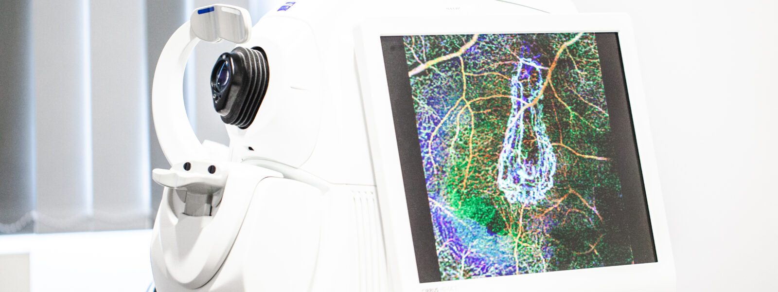
Optical coherence tomography, or OCT of the optic disc, is a laser-based examination of the optic disc. With the help of this examination, we can precisely measure the optic nerve and the surrounding nerve fibers of the retina (retinal nerve fiber layer, RNFL for short). If this examination takes place regularly, a so-called progress assessment can follow.
The values of each individual measurement are compared with values from a norm database. This allows us to make an individual risk analysis and say whether glaucoma, or glaucoma, is present. In addition, a measurement of a specific layer of the macula, ganglion cell thickness, can be measured using optical coherence tomography. It is of great importance in the detection and follow-up of early glaucoma damage.
OCT angiography is a special examination. It allows visualization of the vessels of the optic disc and the vascular density of the surrounding retinal nerve fiber layer. In contrast to vascular imaging of the ocular fundus with fluorescein angiography, this examination is performed without administration of a dye.
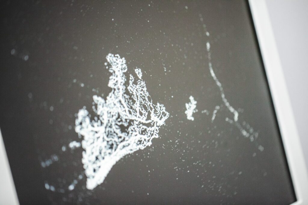
OCT angiography measurements allow us to make statements about the blood flow to your optic disc and inner retinal layers. In particular, the comparison of several images over time then allows an assessment of the stability of the findings.
You would like to know more about the topic or make an appointment? Then simply call us at 0221/871050 or conveniently arrange your desired appointment online.